Journal of Neurology, Neurological Science and Disorders
Metastic Mesothelioma to the Brain: A Potential Differential Diagnosic Challenge
Dalwadi VD, Sheikhi LE, Braun KL, Quist KD and Prayson RA*
Cite this as
Dalwadi VD, Sheikhi LE, Braun KL, Quist KD, Prayson RA (2017) Metastic Mesothelioma to the Brain: A Potential Differential Diagnosic Challenge. J Neurol Neurol Sci Disord 3(1): 025-027. DOI: 10.17352/jnnsd.000016Background: Malignant mesothelioma is a rare neoplasm arising from the mesothelial surfaces of the pleural cavity, peritoneal cavity, tunica vaginalis or pericardium that spreads mainly via direct invasion. While distant metastasis is possible, metastasis to the central nervous system (CNS) is rare.
The Context and Purpose of the Study: We describe a 66-year-old male with a past medical history of mesothelioma, diagnosed approximately 1.5 years prior and treated with resection and chemotherapy, who developed an episode of right facial twitching and was found to have two left cortical frontal masses. After undergoing a frontoparietal craniotomy, the masses were diagnosed as a metastatic mesothelioma.
Main Findings: The tumor was characterized by disordered sheets of large, rounded atypical tumor cells with abundant eosinophilic cytoplasm, irregularly shaped nuclei and prominent nucleoli. Prominent mitotic activity and areas of geographic necrosis were also observed. The tumor demonstrated diffuse positive staining with antibody to calretinin, and focal positive staining with antibodies to D2-40 and WT-1.
Conclusion: Cerebral metastasis of malignant pleural mesothelioma is a rare, but identifiable cause of neurologic symptoms. Specific immunomarkers including calretinin, WT-1 and D2-40 can help distinguish these tumors from more commonly encountered metastases (carcinomas and melanomas)
Abbreviations
Malignant pleural mesothelioma (MPM); Central nervous system (CNS); Computed tomography (CT); Positron emission tomography (PET); Magnetic resonance imaging (MRI)
Background
Malignant mesothelioma is a rare neoplasm arising from the mesothelial surfaces of the pleural cavity, peritoneal cavity, tunica vaginalis or pericardium. The most common type, malignant pleural mesothelioma (MPM), is associated with high mortality and occupational asbestos exposure, with history of exposure identified in approximately 80% of cases [1,2]. The estimated annual incidence of mesothelioma in the United States is approximately 3300 cases per year [3]. The incidence of malignant mesothelioma peaked in 1995, coinciding with diminishing occupational asbestos exposure [2,4]. MPM disease progression is primarily local and constitutes the major cause of death. While distant metastasis is possible via hematogenous spread, metastasis to the central nervous system (CNS) is rare [5]. Herein, we present a case of mesothelioma metastasis to the brain.
Case Presentation
A 66-year-old male with no significant comorbidities presented to his primary care provider with a 7 month history of persistent cough, occasional dysphagia, hoarseness of voice and a 15 pound weight loss. Initial chest x-ray showed multiple pleural-based masses.
Subsequent computed tomography (CT) scan of the chest showed a large left upper lobe, pleural-based mass measuring 43 x 35 mm, a large left lower lobe mass (36 x 26 mm), multiple pleural-based masses and left pleural effusion. Pleural biopsy revealed malignant mesothelioma, biphasic type with positive immunostaining with antibodies to WT-1 (1:100 dilution, DAKO, Carpinteria, CA) and calretinin (1:40 dilution, ThermoFisher, Waltham, MA). He was not considered a good surgical candidate given the extent of disease. A positron emission tomography (PET) scan revealed a large anterior mediastinal hypermetabolic mass. He completed 6 cycles of pemetrexed/carboplatin and continued on maintenance pemetrexed therapy.
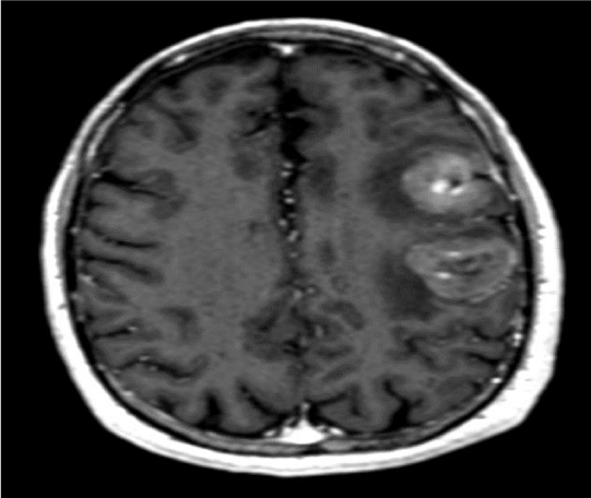
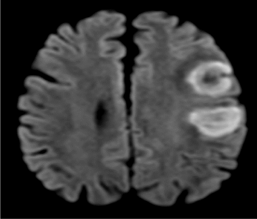
Eighteen months following his diagnosis of mesothelioma, he presented with one-week history of slurred speech, right facial droop and right facial twitching. Magnetic resonance imaging (MRI) of the brain revealed two frontal lesions with local mass effect and vasogenic edema, one in the middle frontal gyrus, measuring 2.1 x 2.5 x 2.8 cm which was T1 hypointense, T2 heterogeneously hypointense, with homogenous enhancement on the postcontrast images, and the second in the left precentral gyrus lesion measuring 2.7 x 3.0 x 2.9 cm (Figures 1,2).
He underwent a left frontoparietal craniotomy for resection of left frontal and left parietal tumors via a single incision and craniotomy. A multidisciplinary brain tumor conference recommended postoperative fractionated gamma knife radiosurgery. The patient was alive at most recent follow-up, three months after surgery, with the intent of following the patient every three months with imaging.
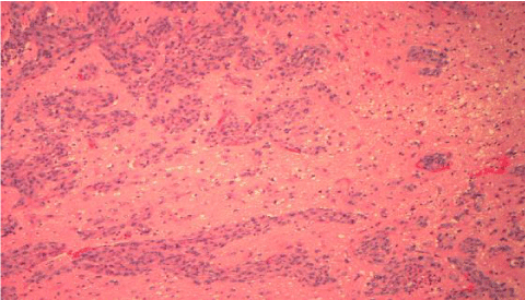
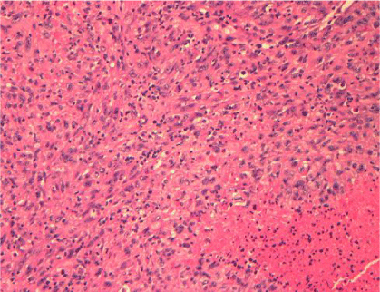
Tissue from the left parietal and frontal lobes was examined. The bulk of the resected tissue represented tumor which was sharply demarcated from the surrounding reactive brain parenchyma (Figure 3). The tumor was characterized by disordered sheets of large rounded atypical tumor cells with abundant eosinophilic cytoplasm, irregularly shaped nuclei and prominent nucleoli (Figure 4). In some areas, the tumor cells assumed a more spindled appearance. There was no discernible gland formation, keratinization or melanin pigment observed. Prominent mitotic activity in excess of 20 mitotic figures in 10 high power microscopic fields was observed. Areas of geographic necrosis were also evident. The tumor demonstrated diffuse positive staining with antibody to calretinin (1:40 dilution; Thermo Fisher Scientific, Wastham, MA) (Figure 5). Additional focal positive staining was seen with antibodies to D2-40 (1:50 dilution, Covance, San Diego, CA) and WT-1. (1:100 dilution; DAKO, Carpinteria, CA) The tumor did not stain with antibodies to melan A (1:40 dilution, Biogenex, Freemont, CA), TTF-1 (prediluted, Ventana, Tucson, AZ), and CEA (1:2000 dilution, DAKO).
Discussion
Malignant mesothelioma is a locally invasive and aggressive tumor of the serosal surfaces primarily associated with occupational asbestos exposure. The prevalence of brain metastasis from MPM, based on a fairly recent retrospective study, is approximately 3% [1,4]. In the review of 171 patients with malignant pleural mesothelioma at autopsy, 4% of patients had distant metastases. The sites most commonly affected were the liver, adrenal glands and kidneys (56, 31 and 30%, respectively). Cerebral metastasis was found in only 3%, involving parietal, frontal, temporal, and cerebellar regions in descending incidence [1,6]. Wronski & Burt concluded that cerebral metastasis at the time of death occurs probably in the range of 5–10% [7]. Metastatic mesothelioma is considered highly aggressive, with less than 5% of patients surviving over 5 years and the median survival reported between 6-12 months [2]. Follow-up in such patients typically involves a multidisciplinary team approach, given the myriad of management considerations.
In our case study, imaging performed prior to resection showed restricted diffusion of the lesions, in addition to vasogenic edema and homogeneous enhancement (Figure 2). The differential for restricted diffusion most commonly includes acute infarcts and abscesses, but a case series of cerebral metastases from a heterogeneous group of source cancers with histopathologic correlation identified this radiologic feature in 7% of metastatic lesions [8]. The most common source tumor was lung carcinoma (37 of 87 patients, 48.6%). This highlights a radiographic feature that assists with the differential of intracranial masses.
As with the current case, a known history of mesothelioma heightens the awareness of the pathologist who will be evaluating the excised tumor and appropriate antibody stains can be applied to confirm a diagnosis. More challenging are cases in which there is no known primary. Morphologically, the tumor often resembles a metastatic large cell carcinoma or melanoma, both much more commonly encountered metastatic neoplasm. Especially for tumors which do not stain with conventional markers of melanoma (melan A, SOX 10 or S-100 protein) or carcinoma (cytokeratin markers or markers which target subsets of adenocarcinomas such as TTF-1, CEA, estrogen/progesterone receptors, PSA, CDX2), consideration of mesothelioma as a possible diagnosis and evaluation with immunomarkers that generally target this tumor (calretinin, WT-1 and D2-40) may be warranted [9]. Also to be considered in the differential diagnosis would be an epithelioid glioblastoma. These tumors usually have focal areas which resemble more typical appearing glioblastoma and would likely stain, at least focally, with GFAP antibody.
Conclusion
Cerebral metastasis of MPM is a rare, but identifiable cause of neurologic symptoms. A prior clinical history is helpful to direct the histologic evaluation. Specific immunomarkers, including calretinin, WT-1 and D2-40, can help distinguish these from more commonly encountered tumors.
- Yamagishi T, Fujimoto N, Miyamoto Y, Asano M, Fuchimoto Y, et al. (2016) Brain Metastases in malignant pleural mesothelioma. Clin Exp Metastasis 33: 231-237. Link: https://goo.gl/73Htzs
- Patel SC, Dowell JE (2016) Modern management of malignant pleural mesothelioma. Lung Cancer: Targets and Therapy 7: 63–72. Link: https://goo.gl/DMrZyk
- Teta MJ, Mink PJ, Lau E, Bonnielin KS, Foster ED. (2008) US mesothelioma patterns 1973-2002: indicators of change and insights into background rates. Eur J Cancer Prev. Link: https://goo.gl/HXfNCX
- Howlader N, Noone AM, Krapcho M, Garshell J, Neyman N, et al. (2013) Surveillance, Epidemiology, and End Results (SEER) Cancer Statistics Review (1975–2010), National Cancer Institute, Bethesda MD, Link: https://goo.gl/mMQEvP
- Miller AC, Miettinen M, Schrump DS, Hassan R (2014) Malignant Mesothelioma and Central Nervous System Metastases. Report of Two Cases, Pooled Analysis, and Systematic Review. Annals of the American Thoracic Society 11: 1075–1081. Link: https://goo.gl/tMYYe6
- Falconieri G, Grandi G, DiBonito L, Bonifacio GD, Giarelli L (1991) Intracranial metastases from malignant pleural mesothelioma. Report of three autopsy cases and review of the literature. Arch Pathol Lab Med 115: 591–595. Link: https://goo.gl/ZBpYjh
- Wronski M, Burt M (1993) cerebral metastases in pleural mesothelioma: case report and review of the literature. J Neurooncol 17: 21–26. Link: https://goo.gl/NnzQXr
- Duygulu G, Yilmaz O, Çalli C, Kitis Ö, Yünten N, et al. (2010) Intracerebral metastasis showing restricted diffusion: Correlation with histopathologic findings. European Journal of Radiology 74: 117-120. Link: https://goo.gl/SK1Y3p
- Ordonez NG (2003) The immunohistochemical diagnosis of mesothelioma: a comparative study of epithelioid mesothelioma and lung adenocarcinoma. Am J Surg Pathol 27: 1031-1051. Link: https://goo.gl/1hUvA6
Article Alerts
Subscribe to our articles alerts and stay tuned.
 This work is licensed under a Creative Commons Attribution 4.0 International License.


 Save to Mendeley
